Details

Electroanatomical Mapping
An Atlas for Clinicians1. Aufl.
|
91,99 € |
|
| Verlag: | Wiley-Blackwell |
| Format: | EPUB |
| Veröffentl.: | 26.08.2011 |
| ISBN/EAN: | 9781444357356 |
| Sprache: | englisch |
| Anzahl Seiten: | 280 |
DRM-geschütztes eBook, Sie benötigen z.B. Adobe Digital Editions und eine Adobe ID zum Lesen.
Beschreibungen
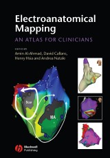
Catheter ablation has become a mainstay in the therapy of cardiac arrhythmias. The development of electroanatomical mapping technologies (such as CARTO) has facilitated more complex ablation procedures. <br /> <p><br /> </p> <p>This brand new book encompasses cardiac arrhythmias and practical tips for users of electroanatomical mapping, providing a color atlas of different arrhythmias, presented as cases, that have been carefully mapped and correlated with clinical and electrogram data.<br /> </p> <p><br /> </p> <p>Including maps from all the major mapping systems such as CARTO, NAVX, ESI, RPM as well as activation maps and voltage maps, this book is an ideal reference book and learning tool for electrophysiologists, electrophysiology fellows and electrophysiology laboratory staff.</p>
Foreword: Dr Marchlinski. <p>1 Electroanatomical mapping technologies: Jeff Hsing (Stanford University Medical Center), Paul J. Wang (Stanford University Medical Center), Amin Al-Ahmad (Stanford University Medical Center).</p> <p>2 Electroanatomical mapping for supraventricular tachycardias: Pirooz Mofrad (Stanford University Medical Center), Amin Al-Ahmad (Stanford University Medical Center), Andrea Natale (Stanford University).</p> <p>3 The utility of electroanatomical mapping in catheter ablation of ventricular tachycardias: Daejoon Anh (Stanford University), Henry H. Hsia (Stanford University Medical Center), David J. Callans (University of Pennsylvania Health System).</p> <p><b>Part I SVT cases.</b></p> <p>4 Electroanatomical mapping for AV nodal reentrant tachycardia using CARTOTM: Henry A. Chen (Stanford University Medical Center), Henry H. Hsia (Stanford University Medical Center).</p> <p>6 Slow pathway ablation: Robert Schweikert (Cleveland Clinic), Luigi Di Biase (Cleveland Clinic and University of Foggia), Andrea Natale (Stanford University).</p> <p>7 Electroanatomical mapping for right-sided accessory pathway: Henry A. Chen (Stanford University Medical Center), Amin Al-Ahmad (Stanford University Medical Center).</p> <p>8 Ablation of an accessory pathway: endocardial and epicardial mapping: Robert Schweikert (Cleveland Clinic), Luigi Di Biase (Cleveland Clinic and University of Foggia), Andrea Natale (Stanford University).</p> <p>9 Focal atrial tachycardia: Daejoon Anh (Stanford University), Henry H. Hsia (Stanford University Medical Center).</p> <p>10 Electroanatomical mapping for right para-hisian atrial tachycardia: Rupa Bala (University of Pennsylvania Health System).</p> <p>11 Right atrial tachycardia after ablation for inappropriate sinus tachycardia: David Callans (University of Pennsylvania Health System).</p> <p>12 Inappropriate sinus tachycardia: Greg Feld (University of California San Diego Medical Center).</p> <p>13 Focal left atrial tachycardia: Sumeet K. Mainigi (Albert Einstein Medical Center).</p> <p>14 Electroanatomic mapping of right atrial tricuspid annulus atrial tachycardia: Pirooz Mofrad (Stanford University Medical Center), Henry H. Hsia (Stanford University Medical Center).</p> <p>15 Electroanatomical mapping of a left-sided atrial tachycardia: Kevin J. Makati (Tufts University-New England Medical Center), N. A. Mark Estes III (Tufts University School of Medicine).</p> <p>16 Focal atrial tachycardia: John Triedman (Children’s Hospital Boston).</p> <p>17 Ectopic atrial tachycardia: George Van Hare (Stanford University).</p> <p>18 Electroanatomical mapping for scar-based reentrant atrial tachycardia: Pirooz Mofrad (Stanford University Medical Center), Amin Al-Ahmad (Stanford University Medical Center).</p> <p>19 Atrial fl utter in a patient post-Fontan: Riccardo Cappato (IRCCS Policlinico San Donato).</p> <p>20 Recurrent atrial flutter after isthmus ablation: David Callans (University of Pennsylvania Health System).</p> <p>21 Scar-based reentrant atrial tachycardia in a patient with congenital heart disease: Ricarrdo Capato (IRCCS Policlinico San Donato).</p> <p>22 A case of post-Maze atrial fl utter: Tamer S. Fahmy (Cairo University Hospitals), Andrea Natale (Stanford University).</p> <p>23 A case of atypical atrial fl utter: Tamer Fahmy (Cairo University Hospitals), Dimpi Patel (Cleveland Clinic), Andrea Natale (Stanford University).</p> <p>24 Spontaneous scar-based atypical and typical AFL: Greg Feld (University of California San Diego Medical Center).</p> <p>25 Left atrial flutter after pulmonary vein isolation: Richard Hongo (Marin General Hospital), Andrea Natale (Stanford University).</p> <p>26 Scar-based reentrant atrial tachycardia: Henry Hsia (Stanford University Medical Center).</p> <p>27 Atypical atrial flutter following circumferential left atrial ablation: James David Allred (University of Alabama at Birmingham), Harish Doppalapudi (University of Alabama at Birmingham), G. Neal Kay (University of Alabama at Birmingham).</p> <p>28 Scar-related intraatrial reentrant tachycardia: Kevin J. Makati (Tufts University-New England Medical Center), N. A. Mark Estes III (Tufts University School of Medicine).</p> <p>29 Electroanatomic mapping for atrial flutter: Pirooz Mofrad (Stanford University Medical Center), Amin Al-Ahmad (Stanford University Medical Center).</p> <p>30 Electroanatomical mapping for incessant small macroreentrant left atrial tachycardia following catheter ablation of atrial fibrillation:.</p> <p>Hiroshi Nakagawa (University of Oklahoma Health Sciences Centre), Warren Jackman (University of Oklahoma Health Sciences Center).</p> <p>31 Macroreentry left atrial tachycardia: Linda Huffer (Walter Reed Army Medical Center), William Stevenson (Harvard Medical School).</p> <p>32 Focal atrial tachycardia in a patient with a Fontan: John Triedman (Children’s Hospital Boston).</p> <p>33 Macrareentrant atrial tachycardia in patient with a history of tetralogy of Fallot: John Triedman (Children’s Hospital Boston).</p> <p>34 Double loop macroreentrant atrial tachycardia in a patient with tetralogy of Fallot: John Triedman (Children’s Hospital Boston).</p> <p>35 Atrial flutter in a patient post-Fontan: John Triedman (Children’s Hospital Boston).</p> <p>36 Atrial tachycardia in a patient post-Mustard procedure: John Triedman (Children’s Hospital Boston).</p> <p>37 Repeat interruption of ongoing atrial fibrillation during RF pulse delivery: Riccardo Cappato (IRCCS Policlinico San Donato).</p> <p><b>Part II VT cases.</b></p> <p>38 PVC and nonsustained ventricular tachycardia baltion in a child: George Van Hare (Lucile Packard Children’s Hospital).</p> <p>39 Electroanatomical mapping for ventricular premature complexes from the right ventricular outflow tract: Henry A. Chen (Stanford University Medical Center), Paul J. Wang (Stanford University School of Medicine).</p> <p>40 Ablation of idiopathic RV ventricular tachycardia in an unusual location using EnSite mapping: David Callans (University of Pennsylvania Health System).</p> <p>41 Ablation of poorly inducible fascicular ventricular tachycardia: Jason Jacobson (Department of Medicine at Northwestern University).</p> <p>42 Electroanatomic mapping for left anterior fascicular ventricular tachycardia: David Callans (University of Pennsylvania Health System).</p> <p>43 Left ventricular tachycardia originating in basal diverticulum: Sumeet K. Mainigi (Albert Einstein Medical Center).</p> <p>44 PVC originating near aortic cusps: Sumeet K. Mainigi (Albert Einstein Medical Center).</p> <p>45 Right ventricular outflow tachycardia: Kevin J. Makati, (Tufts University-New England Medical Center) N. A. Mark Estes III (Tufts University School of Medicine).</p> <p>46 Right ventricular outflow tract polymorphic ventricular tachycardia: Luis C. Sáenz (Fundacion Cardio Infantil-Instituto de Cardiologia), Miguel A. Vacca (Fundación Cardioinfantil, Instituto de Cardiología), Andrea Natale (Stanford University).</p> <p>47 Left aortic cusp ventricular tachycardia: John Sussman (Morristown Memorial Hospital).</p> <p>48 Ventricular tachycardia in patient with tetralogy of Fallot: Dajoon Anh (Stanford University), Amin Al-Ahmad (Stanford University Medical Center), Henry Hsia (Stanford University Medical Center).</p> <p>49 Ventricular tachycardia in a patient with cardiac sarcoid: Rupa Bala (University of Pennsylvania Health System), David J. Callans (University of Pennsylvania Health System).</p> <p>50 Ventricular tachycardia in an area of left ventricular noncompaction: John Sussman (Morristown Memorial Hospital).</p> <p>51 Ventricular tachycardia in a patient with arrhythmogenic right ventricular dysplasia: Rupa Bala (University of Pennsylvania Health System).</p> <p>52 Electroanatomical mapping for arrhythmogenic right ventricular dysplasia: Henry Hsia (Stanford University Medical Center).</p> <p>53 Epicardial mapping and ablation of nonischemic ventricular tachycardia: James David Allred (University of Alabama at Birmingham), Harish Doppalapudi (University of Alabama at Birmingham), G. Neal Kay (University of Alabama at Birmingham).</p> <p>54 Double-outlet right ventricle ventricular tachycardia: James David Allred (University of Alabama at Birmingham), Takumi Yamada (University of Alabama at Birmingham), Harish Doppalapudi (University of Alabama at Birmingham), Yung R. Lau (University of Alabama at Birmingham), G. Neal Kay (University of Alabama at Birmingham).</p> <p>55 Voltage mapping of the right ventricle in arrythmogenic right ventricular dysplasia: Kevin J. Makati (Tufts University-New England Medical Center), N. A. Mark Estes III (Tufts University School of Medicine).</p> <p>56 Ventricular tachycardia related to arrhythmogenic right ventricular dysplasia: Luis C. Sáenz (Fundacion Cardio Infantil-Instituto de Cardiologia), Miguel A. Vacca (Fundación Cardioinfantil, Instituto de Cardiología), Andrea Natale (Stanford University).</p> <p>57 Electroanatomic mapping for scar-mediated right ventricular tachycardia: Linda Huffer (Walter Reed Army Medical Center), William Stevenson (Harvard Medical School).</p> <p>58 Post-myocardial infarction ventricular tachycardia: John Sussman (Morristown Memorial Hospital).</p> <p>59 Endocardial and epicardial ventricular tachycardia ablation: Daejoon Anh (Stanford University), Paul C. Zei (Stanford University Medical Center), Henry Hsia (Stanford University Medical Center).</p> <p>60 Ablation of ventricular tachycardia in the setting of coronary artery disease using dynamic substrate mapping: David Callans (University of Pennsylvania Health System).</p> <p>61 Endocardial and epicardial mapping for ischemic ventricular tachycardia: Tamer S. Fahmy (Cairo University Hospitals), Oussama M. Wazni (Cleveland Clinic), Moataz Ali (Cairo University and Cleveland Clinic), Robert A. Schweikert (Cleveland Clinic), Andrea Natale (Stanford University).</p> <p>62 Electroanatomical mapping for ischemic ventricular tachycardia: Henry Hsia (Stanford University Medical Center).</p> <p>63 Electroanatomical mapping for scar-based reentrant ventricular tachycardia: Henry A. Chen (Stanford University Medical Center), Henry H. Hsia (Stanford University Medical Center).</p> <p>64 Electroanatomical mapping for scar-based reentrant ventricular tachycardia:Henry A. Chen (Stanford University Medical Center), Henry H. Hsia (Stanford University Medical Center).</p> <p>65 Epicardial mapping and ablation of ischemic ventricular tachycardia: James David Allred (University of Alabama at Birmingham), Harish Doppalapudi (University of Alabama at Birmingham), G. Neal Kay (University of Alabama at Birmingham).</p> <p>66 Substrate modifi cation in hemodynamically unstable infarct-related ventricular tachycardia: Kevin J. Makati (Tufts University-New England Medical Center) , N. A. Mark Estes III (Tufts University School of Medicine).</p> <p>67 Electroanatomic mapping for scar-mediated left ventricular tachycardia: Linda Huffer (Walter Reed Army Medical Center), William Stevenson (Harvard Medical School).</p> <p>68 Ventricular tachycardia, endocardial and epicardial mapping: Jonathan Sussman (Morristown Memorial Hospital).</p> <p>69 Endocardial and epicardial mapping for ventricular tachycardia in the setting of myocarditis: David Callans (University of Pennsylvania Health System).</p> <p>70 Multiple left ventricular basal ventricular tachycardias in a patient with dilated cardiomyopathy: Jason Jacobsen (Department of Medicine at Northwestern University).</p> <p>71 Epicardial ventricular tachycardia in a patient with nonischemic cardiomyopathy: Rupa Bala (University of Pennsylvania Health System).</p> <p><b>Part III Tips and tricks.</b></p> <p>72 Three-dimensional mapping and navigation with the EnSite Array and NavX system: Craig A. Swygman (Providence St. Vincent Hospital), Blair D. Halperin (Providence St. Vincent Hospital).</p> <p>73 CARTO XP: Tips and tricks: William (Marty) Castell (Testamur NASPExAM AP/EP).</p> <p>Index.</p>
<b>Amin Al-Ahmad, MD</b><br /> Cardiac electrophysiologist<br /> Stanford University Medical Center<br /> Stanford, CA, USA<br /> <p><b>David Callans, MD</b><br /> Professor of Medicine<br /> Director of the Electrophysiology Laboratory<br /> University of Pennsylvania<br /> Philadelphia, PA, USA<br /> </p> <p><b>Henry H. Hsia, MD</b><br /> Associate Professor of Medicine<br /> Stanford University<br /> Stanford, CA, USA<br /> </p> <p><b>Andrea Natale, MD<br /> </b>Department of Cardiology<br /> Stanford University Medical Center<br /> Stanford, CA, USA</p>
Catheter ablation has become a mainstay in the therapy of cardiac arrhythmias. The development of electroanatomical mapping technologies (such as CARTO) has facilitated more complex ablation procedures. <p>This brand new book encompasses cardiac arrhythmias and practical tips for users of electroanatomical mapping, providing a color atlas of different arrhythmias, presented as cases, that have been carefully mapped and correlated with clinical and electrogram data.</p> <p>Including maps from all the major mapping systems such as CARTO, NAVX, ESI, RPM as well as activation maps and voltage maps, this book is an ideal reference book and learning tool for electrophysiologists, electrophysiology fellows and electrophysiology laboratory staff.</p>


