Details

Veterinary Head and Neck Imaging
1. Aufl.
|
174,99 € |
|
| Verlag: | Wiley-Blackwell |
| Format: | |
| Veröffentl.: | 15.12.2021 |
| ISBN/EAN: | 9781119118626 |
| Sprache: | englisch |
| Anzahl Seiten: | 800 |
DRM-geschütztes eBook, Sie benötigen z.B. Adobe Digital Editions und eine Adobe ID zum Lesen.
Beschreibungen
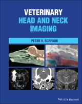
<p><b>A complete, all-in-one resource for head and neck imaging in dogs, cats, and horses </b></p> <p><i>Veterinary Head and Neck Imaging </i>is a comprehensive reference for the diagnostic imaging of the head and neck in dogs, cats, and horses. The book provides a multimodality, comparative approach to neuromusculoskeletal, splanchnic, and sense organ imaging. It thoroughly covers the underlying morphology of the head and neck and offers an integrated approach to understanding image interpretation.</p> <p>Each chapter covers a different area and discusses developmental anatomy, gross anatomy, and imaging anatomy, as well as the physical limitations of different modalities and functional imaging. Commonly encountered diseases are covered at length.</p> <p><i>Veterinary Head and Neck Imaging</i> includes all relevant information from each modality and discusses multi-modality approaches. The book also includes:</p> <ul> <li>A thorough introduction to the principles of veterinary head and neck imaging, including imaging technology, interpretation principles, and the anatomic organization of the head and neck</li> <li>Comprehensive explorations of musculoskeletal system and intervertebral disk imaging, including discussions of degenerative diseases, inflammation, and diskospondylitis</li> <li>Practical discussions of brain, spinal cord, and cerebrospinal fluid and meninges imaging, including discussions of trauma, vascular, and neoplastic diseases</li> <li>In-depth treatments of peripheral nerve, arterial, venous and lymphatic, respiratory, and digestive system imaging</li> </ul> <p><i>Veterinary Head and Neck Imaging </i>is a must-have resource for veterinary imaging specialists and veterinary neurologists, as well as for general veterinary practitioners with a particular interest in head and neck imaging.</p>
<p>Preface xi</p> <p><b>Section 1 Introduction to Head and Neck Imaging in Animals 1</b></p> <p><b>1 Some Basic Concepts About Head and Neck Anatomy 3</b></p> <p>1.1 Terms of Location, Orientation, and Movement 4</p> <p>1.2 External Features of the Head and Neck 11</p> <p>1.3 Overview of Neuroanatomic Localization During Neuroimaging 13</p> <p>1.3.1 Divisions of the Central Nervous System 15</p> <p>1.3.2 Neuroaxis Localization 20</p> <p>1.3.3 Clinical Descriptors for the Location of Intracranial Abnormalities 26</p> <p>References 32</p> <p><b>2 Some Basic Concepts about Medical Imaging 33</b></p> <p>2.1 Introduction 33</p> <p>2.1.1 What is an Image? 33</p> <p>2.1.2 What is Medical Imaging? 34</p> <p>2.2 Medical Imaging Devices 39</p> <p>2.2.1 Imaging Technologies 39</p> <p>2.2.2 Imaging Techniques, Applications, and Examinations 41</p> <p>2.3 The Medical Image 47</p> <p>2.3.1 Picture Elements and Volumetric Picture Elements 47</p> <p>2.3.2 Representing Tissue Characteristics through the Grayscale 50</p> <p>2.3.3 Resolution 52</p> <p>2.4 Image Evaluation 56</p> <p>2.4.1 Getting Started 56</p> <p>2.4.2 Imaging Signs and Patterns 59</p> <p>2.4.3 Image Evaluation 66</p> <p>References 67</p> <p><b>Section 2 Musculoskeletal Imaging 69</b></p> <p><b>3 The Musculoskeletal System 71</b></p> <p>3.1 Imaging Anatomy 71</p> <p>3.1.1 Bone 71</p> <p>3.1.2 Imaging Anatomy – Joints and Ligaments 90</p> <p>3.1.3 Muscle and Tendons 96</p> <p>3.1.3.1 Fascia and Fascial Compartments 111</p> <p>3.2 Musculoskeletal Abnormalities 119</p> <p>3.2.1 Developmental Malformations 120</p> <p>3.2.1.1 Cranium, Face, and Craniocervical Junction 120</p> <p>3.2.1.2 Vertebrae 126</p> <p>3.2.2 Degenerative Diseases 131</p> <p>3.2.2.1 Joints 132</p> <p>3.2.2.2 Vertebrae 138</p> <p>3.2.3 Inflammatory Diseases 138</p> <p>3.2.3.1 Infectious 142</p> <p>3.2.3.2 Noninfectious 146</p> <p>3.2.4 Neoplasia 150</p> <p>3.2.5 Nutritional, Metabolic, Toxic Diseases 162</p> <p>3.2.6 Trauma 170</p> <p>3.2.6.1 Soft-tissue Trauma 171</p> <p>3.2.6.2 Fracture 173</p> <p>3.2.6.3 Dislocation 183</p> <p>References 190</p> <p><b>4 Intervertebral Disks 198</b></p> <p>4.1 Imaging Anatomy 198</p> <p>4.2 Intervertebral Disk Abnormalities 200</p> <p>4.2.1 Developmental Malformations 200</p> <p>4.2.2 Infection/Inflammation 202</p> <p>4.2.3 Trauma 204</p> <p>4.2.4 Degeneration 208</p> <p>4.2.5 Herniation 214</p> <p>References 236</p> <p>Section 3 Nervous System Imaging 241</p> <p><b>5 Cerebrospinal Fluid 243</b></p> <p>5.1 Imaging Anatomy 243</p> <p>5.2 CSF Production, Absorption, and Flow 246</p> <p>5.3 Cerebrospinal Fluid Abnormalities 250</p> <p>5.3.1 Intra-Axial Fluid Accumulations 251</p> <p>5.3.2 Extra-Axial Fluid Accumulations 268</p> <p>5.3.3 Intramedullary Fluid Accumulations 272</p> <p>5.3.4 Extramedullary Fluid Accumulations 275</p> <p>References 285</p> <p><b>6 The Central Nervous System 289</b></p> <p>6.1 Imaging Anatomy 289</p> <p>6.2 Brain and Spinal-Cord Abnormalities 297</p> <p>6.2.1 Imaging Patterns of CNS Disease 297</p> <p>6.2.1.1 Some Additional Imaging Signs 300</p> <p>6.2.1.2 Contrast Enhancement 302</p> <p>6.2.2 Secondary Intracranial Abnormalities 308</p> <p>6.2.2.1 Intracranial Hypertension 308</p> <p>6.2.2.2 Cerebral Edema 310</p> <p>6.2.2.3 MRI Signs Induced by Seizures 313</p> <p>6.2.2.4 Brain Herniation 314</p> <p>6.2.3 Developmental Malformations 321</p> <p>6.2.4 Vascular Disorders 328</p> <p>6.2.4.1 Ischemia 329</p> <p>6.2.4.2 Hemorrhage 341</p> <p>6.2.4.3 Hemorrhagic Infarction 349</p> <p>6.2.5 Trauma 349</p> <p>6.2.5.1 Traumatic Brain Injury 350</p> <p>6.2.5.2 Traumatic Spinal-Cord Injury 356</p> <p>6.2.6 Neoplasia 364</p> <p>6.2.7 Inflammatory Diseases 385</p> <p>6.2.7.1 Infectious 400</p> <p>6.2.7.2 Noninfectious 428</p> <p>6.2.8 Degenerative Diseases 440</p> <p>References 452</p> <p><b>7 The Peripheral Nervous System 475</b></p> <p>7.1 Imaging Anatomy 475</p> <p>7.1.1 Cranial Nerves 478</p> <p>7.1.2 Spinal Nerves 495</p> <p>7.1.2.1 The Cervical Nerves 499</p> <p>7.1.2.2 The Brachial Plexus 500</p> <p>7.1.2.3 The Sympathetic Division 501</p> <p>7.2 Peripheral Nerve Abnormalities 503</p> <p>7.2.1 Neoplasia 506</p> <p>7.2.2 Trauma 510</p> <p>7.2.3 Inflammatory Diseases 511</p> <p>7.2.4 Compression 512</p> <p>7.2.5 Degenerative Diseases 513</p> <p>References 522</p> <p>Section 4 Splanchnic (Viscera), Vascular, and Sense Organ Imaging 525</p> <p><b>8 The Digestive System 527</b></p> <p>8.1 Imaging Anatomy 527</p> <p>8.1.1 Oral Cavity 527</p> <p>8.1.2 Pharynx 540</p> <p>8.1.3 Cervical Esophagus 544</p> <p>8.2 Digestive Track Abnormalities 545</p> <p>8.2.1 Developmental Malformations 545</p> <p>8.2.2 Dysphagia 552</p> <p>8.2.3 Neoplasia 564</p> <p>8.2.4 Inflammation 579</p> <p>References 598</p> <p><b>9 The Respiratory System 602</b></p> <p>9.1 Imaging Anatomy 602</p> <p>9.1.1 Nasal Cavities and External Nose 602</p> <p>9.1.2 Paranasal Sinuses 606</p> <p>9.1.3 Nasopharynx, Larynx, and Cervical Trachea 611</p> <p>9.2 Respiratory Track Abnormalities 620</p> <p>9.2.1 Developmental Anomalies 620</p> <p>9.2.2 Inflammation/Infection 625</p> <p>9.2.3 Neoplasms 630</p> <p>9.2.4 Degenerative Disorders 634</p> <p>References 668</p> <p><b>10 Sense Organs, Circulatory System, and Endocrine System 673</b></p> <p>10.1 Imaging Anatomy 673</p> <p>10.1.1 Eye 673</p> <p>10.1.2 Ear 674</p> <p>10.1.3 Circulatory System 681</p> <p>10.1.4 Endocrine System 691</p> <p>10.2 Orbital Disorders 693</p> <p>10.2.1 Trauma 695</p> <p>10.2.2 Inflammatory Disease 702</p> <p>10.2.3 Neoplasms 705</p> <p>10.3 Ear Disorders 707</p> <p>10.3.1 Ear Diseases 707</p> <p>10.3.2 Guttural Pouch Disease 715</p> <p>10.3.3 Imaging Patterns of Disease 718</p> <p>10.4 Circulatory and Endocrine Disorders 722</p> <p>10.4.1 Developmental Anomalies 722</p> <p>10.4.2 Endocrine Disorders 723</p> <p>10.4.3 Circulatory System Disorders 733</p> <p>References 764</p> <p>Index 773</p>
<P><B>The author</B></P> <P><B>Peter V. Scrivani, DVM, DACVR,</B> is Associate Professor of Veterinary Imaging at Cornell University’s College of Veterinary Medicine in Ithaca, New York, USA.
<p><b>A complete, all-in-one resource for head and neck imaging in dogs, cats, and horses </b></p> <p><i>Veterinary Head and Neck Imaging </i>is a comprehensive reference for the diagnostic imaging of the head and neck in dogs, cats, and horses. The book provides a multimodality, comparative approach to neuromusculoskeletal, splanchnic, and sense organ imaging. It thoroughly covers the underlying morphology of the head and neck and offers an integrated approach to understanding image interpretation.</p> <p>Each chapter covers a different area and discusses developmental anatomy, gross anatomy, and imaging anatomy, as well as the physical limitations of different modalities and functional imaging. Commonly encountered diseases are covered at length.</p> <p><i>Veterinary Head and Neck Imaging</i> includes all relevant information from each modality and discusses multi-modality approaches. The book also includes:</p> <ul> <li>A thorough introduction to the principles of veterinary head and neck imaging, including imaging technology, interpretation principles, and the anatomic organization of the head and neck</li> <li>Comprehensive explorations of musculoskeletal system and intervertebral disk imaging, including discussions of degenerative diseases, inflammation, and diskospondylitis</li> <li>Practical discussions of brain, spinal cord, and cerebrospinal fluid and meninges imaging, including discussions of trauma, vascular, and neoplastic diseases</li> <li>In-depth treatments of peripheral nerve, arterial, venous and lymphatic, respiratory, and digestive system imaging</li> </ul> <p><i>Veterinary Head and Neck Imaging </i>is a must-have resource for veterinary imaging specialists and veterinary neurologists, as well as for general veterinary practitioners with a particular interest in head and neck imaging.</p>
Diese Produkte könnten Sie auch interessieren:

Handbook of Applied Dog Behavior and Training, Procedures and Protocols

von: Steven R. Lindsay

131,99 €


